
Anichstraße 35
6020 Innsbruck
Fax: +43 512 504 27199
Email: Adriano.Crismani@i-med.ac.at
Website: http://www.zmk-innsbruck.at/Kieferorthopaedie.9.0.html
Research year
Research Branch (ÖSTAT Classification)
104024, 302033, 302034, 302044, 302071, 302088, 301108, 211904
Keywords
curve of Spee, Inflammasome, lateral incisors width, lip pressure, NLRP3, oral irrigator, orthodontic mini screws, orthodontic tooth movement, surface-coated materials, and tooth agenesis
Research Focus
- The role of the NLRP3 inflammasome in orthodontic tooth movement (OTM).
- The relationship between the mandibular Curve of Spee (COS) and a persisting primary second mandibular molar (ppM2).
- Evaluation of osseointegration of loaded strontium-functionalised orthodontic mini screws (Ti-Sr-O).
- Evaluation of the standard value of the lateral incisors in the upper jaw in Tyrol.
- Comparison of two home interdental oral hygiene products.
- Lip pressure before, during and after orthodontic treatment.
General Facts
The University Hospital for Orthodontics is part of the Department of Dental and Oral Medicine and Cranio-Maxillofacial and Oral Surgery. There is no extra laboratory personnel.
The major aim of our basic and clinical research activities in the field of orthodontics is to increase knowledge in the respective research areas and to optimise patient treatment.
The current research topics of the University Hospital for Orthodontics focus on the role of the NLRP3 inflammasome in orthodontic tooth movement (OTM), strontium-functionalised orthodontic mini screws, the standard value of the lateral incisors in the upper jaw and the interdental oral hygiene in orthodontic patients. Further clinical research focuses on the bone quality upon orthodontic tooth movement, the analyses of the oral microbiome, the transfer accuracy with indirect bonding using 3D-printed trays and the lip pressure before during and after orthodontic treatment.
We collaborate closely with a number of clinical and scientific units at the Medical University of Innsbruck as well as international research partners. Currently, there are strong collaboration activities on the Innsbruck campus with the University Hospital for Restorative Dentistry and Periodontology , the University Hospital for Cranio-Maxillofacial and Oral Surgery, the University Hospital for Dermatoloy, Venerology and Allergoloy, the University Hospital for Radiology and the Institute of Pathology and Molecular Pathology. For outside collaborations and core facilities, see below.
Research
The NLRP3 Inflammasome and Orthodontic Tooth Movement – A Mouse Model
Lisa Schieffer et al.
This study aims to investigate the role of the NLRP3 inflammasome in orthodontic tooth movement (OTM) and should shed light on the processes involved in bone transformation upon orthodontic treatment. Effects of tooth movement on inflammatory pathways, with special emphasis on the expression of pro-inflammatory cytokines should be clarified. As studies reported a negative co-regulation between physiological oestrogen levels and the speed of OTM gender differences should be evaluated.
NLRP3 depleted mice (NLRP3-/-) and wild-type littermates (WT; males and females) were used in a model of experimental OTM induced by nitinol coil-springs that were bonded between the left first maxillary molar and the upper incisors (Fig. 1).
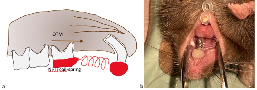
Teeth in the right maxillae served as contralateral controls. After 7, 14, and 21 days of constant orthodontic force, mice were sacrificed, the jaws were removed and analysed by micro-computed tomography (distance for mesial movement of the first molar was measured) and histological analyses (TRAP-staining). To monitor the onset of the inflammasome upon OTM, cytokine levels of IL-1β were measured in blood plasma by enzyme-linked immunosorbent assay (ELISA). Additionally, general health parameters (blood, bodyweight) were analysed.
Micro-computed tomography illustrated that the amount of tooth movement was negligible in NLRP3-/- mice compared to WT mice. Female mice demonstrated little OTM compared to WT male mice. TRAP staining showed statistically significant different more osteoclasts in WT mice in contrast to the NLRP3-/- mice.
ELISA showed low serum levels of IL-1β in all mice, indicating that the inflammation is at least systematically undetectable. Weight and blood analysation gave a survey of good general health and a sufficient nutritional status of the mice.
Orthodontic tooth movement experiments in NLRP3-/- mice discover that the NLRP3 inflammasome is involved in osteoclast induction at the alveolar bone surface on the compression side. This study reveals the possibility that NLRP3 plays a mechanosensory role in OTM. Further, results suggest a negative co-regulation of female hormones in molecular pathways of tooth movement.
Further results, such as bone quality, FACS analyses and analyses of the oral microbiome are still outstanding.
Curve of Spee and Second Mandibular Premolar Agenesis—Provide Knowledge and Future Perspectives
Lisa Schieffer et al.
We investigated the relationship between the mandibular Curve of Spee (COS) and a persisting primary second mandibular molar (ppM2) due to an agenesis of the second mandibular premolar, using a digital software technique.
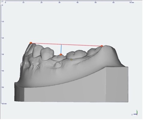
Digital dental casts were obtained from 200 patients at the Department of Orthodontics in Innsbruck and Vienna, Austria. Patients (age-, gender- and malocclusion-matched) were equally divided into two groups (n = 100) according to the existence of a ppM2. COS depth, overjet, overbite and angle-classification were measured digitally using the OnyxCeph3TM (version 3.2.147) software (Fig. 2).
ANOVA and Kruskal–Wallis tests were used to analyse relationships. For statistical analyses, p < 0.05 was considered as statistically significant and p < 0.01 as highly significant. Results were visualised with box plots and bar charts.
The deepest COS was present in patients with a ppM2. Furthermore, a positive correlation was shown between COS depth and angle-class II, between COS depth and age, as well as between COS depth and overbite. No gender differences could be observed.
In our study population, the COS depth was dependent on whether or not there is a ppM2 due to an agenesis of a second mandibular premolar as well as on the malocclusion in sagittal direction.
Osseointegration of Strontium-Coated Loaded Orthodontic Mini Screws Studied by Histology, Synchrotron X-ray Fluorescence and Diffraction Imaging
Natalie Schenz-Spisic et al.
Strontium is known for its antiosteoporotic effects, not only through systemic administration of strontium ranelate, but also through sustained local release on strontium-coated titanium mini screws. The objective of this study was to evaluate the degree of osseointegration via histomorphometry of loaded strontium-functionalised orthodontic mini screws (Ti-Sr-O) and the effect of the Sr coating on the elemental composition of the bone and the coatings effects on bone mineral size and orientation.
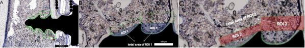
Strontium-coated (Ti-Sr-O) mini screws prepared by a magnetron sputtering process and grade 4 titanium mini screws (Ti) were inserted monocortically into either tibia of 30 male Wistar rats and loaded through a coil spring with a force of 25 cN. After a two-, four- and six-week healing period, bone-to-implant contact (BIC%) and peri-implant bone formation (BF%) in defined regions of interest (ROI-I; ROI-II) were measured on both sides of the mini screws (compression /tension side of the applied force) via histomorphometry (Fig. 3).
Furthermore, samples were investigated at the P06 synchrotron beamline at DESY (Hamburg) to simultaneous measure the atomic composition with X-ray fluorescence (XRF) and the crystallite size/orientation with X-ray diffraction (XRD) used in mapping modes to create multimodal XRF and XRD images.
After two weeks Ti-Sr-O functionalised mini screws showed significantly more bone formation in ROI-I than in the control group with Ti mini screws at the tension (p**=0.005) and the pressure (p*=0.018) side. The histomorphometric analysis four weeks postoperatively showed significantly increased BIC for Ti-Sr-O mini screws at the tension side (p*=0.029). The XRF mapping four weeks postoperatively showed an increased level of Sr/Ca in the peri-implant bone (p**=0.0036). Analysis of the (002) XRD diffraction peak showed a significant change in multiple parameters in the distribution of the degree of orientation (DoO) in the crystallographic c-axis of the mineral crystals as obtained by a linear mixed effects model that considered whether the screws were Sr-coated and whether to Sr/Ca-ratio was high or low.
Sr-functionalised mini screws could serve as a potential modification for temporary anchorage devices in orthodontics with improved early osseointegration and consequently higher success rates.
Evaluation of the Standard Value of the Lateral Incisors in the Upper Jaw in Tyrol (Austria)
Irene Artioli et al.
The maxillary incisors play the most important role in the smile aesthetics. The overall prevalence of peg-shaped lateral incisors is 1.8%. Women are affected 1.35 times more than men. This knowledge is important during the planning of dental treatments in order to achieve an optimal aesthetic result. Aesthetic dentistry tries to find geometric or mathematical rules that describe the proportion of the front teeth.
In this prospective study we want to define a standard value for the lateral incisors width. The findings of this work could also be used for further studies, where the average width of the lateral incisors in the maxilla is required. In addition, the collected data can serve as support for dentists who treat patients with high aesthetical expectations.
The aim of this work is to define the normal and hypoplasia values for the lateral incisors in the upper jaw and to compare the measured values with the available literature.
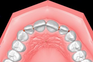
The first 100 consecutive patients at the Department of Dentistry in Innsbruck who meet the inclusion criteria will be included. To measure the teeth, an intraoral scan (iTeroTM) of the maxilla will be performed and the lateral incisors in the upper jaw will be measured (OnyxCeph3TM) (Fig.4).
With this study a standard value for the upper incisor teeth and a ratio between the lateral and the central incisors can be determined. Thus, it is possible to aesthetically rebuild a lateral incisor in case of hypoplasia e.g. after orthodontic treatment.
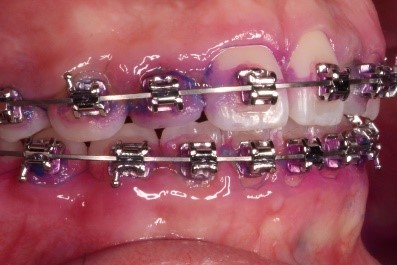
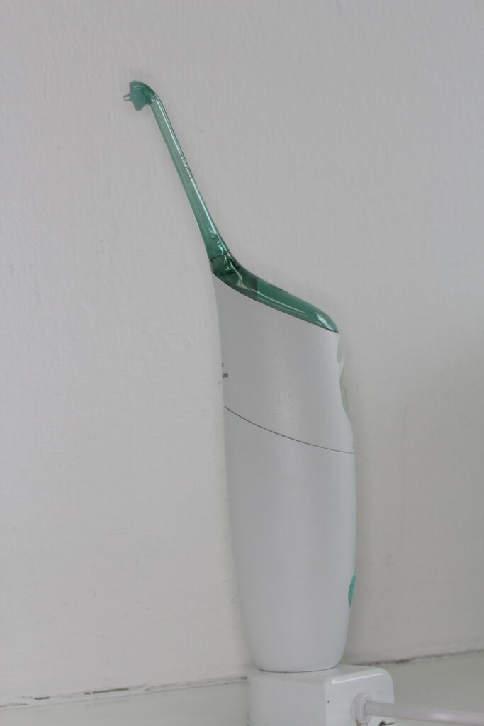
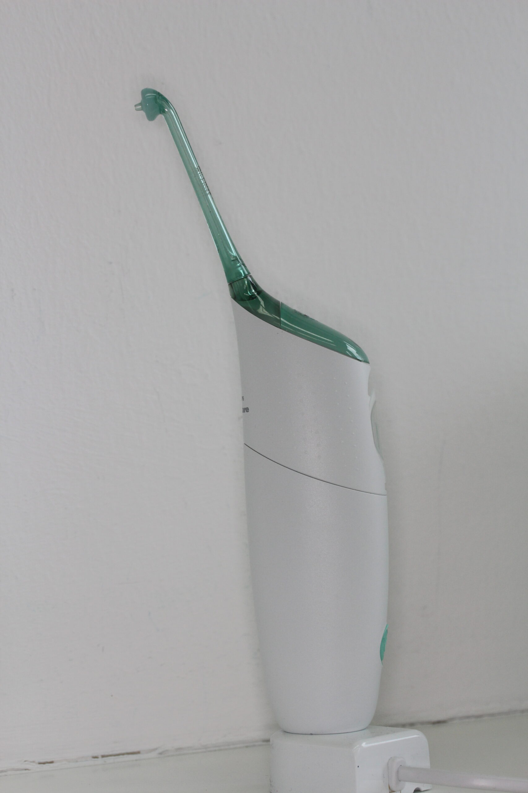
Clinical Study comparing two Home Interdental Oral Hygiene Products in Adolescent Orthodontic Patients
Manuel Kasslatter et al.
Orthodontic patients struggle with interdental cleaning calling for simpler mechanical devices to reduce the high plaque levels. The present study aimed to compare the cleansing efficacy of an oral irrigator with that of dental flossing in patients with fixed braces after four weeks of home-use.
The study design is a randomised and single-blinded cross-over study. After twenty-eight days using the products at home, hygiene indices (Rustogi Modified Navy Plaque Index (RMNPI); Gingival bleeding index (GBI) were compared between test (oral irrigator) and control product (dental floss) (Fig. 5,6).
Seventeen adult test persons completed the study. After 28 days of cleaning with the oral irrigator, RMNPI was 54.96 % (46.91 – 66.05) compared to 52.98 % (42.75 – 65.60) with dental floss (p = 0.029). Subgroup analysis revealed that the higher cleansing efficacy of the dental floss is attributable to buccal and marginal areas. GBI after the test phase with the oral irrigator was 12.96 % (7.14 – 24.31) and statistically significantly higher compared to 8.33 % (5.84 – 15.33) with dental floss (p = 0.030) which could be seen in all subgroups.
Oral irrigators do not remove plaque and reduce gingival bleeding as efficiently as dental floss in easily accessible regions. However, in posterior regions, where the patients struggled with the application of dental floss, the oral irrigator showed similar results.
Oral irrigators should only be recommended to orthodontic patients who are not compliant with dental flossing and interdental brushes are not applicable.
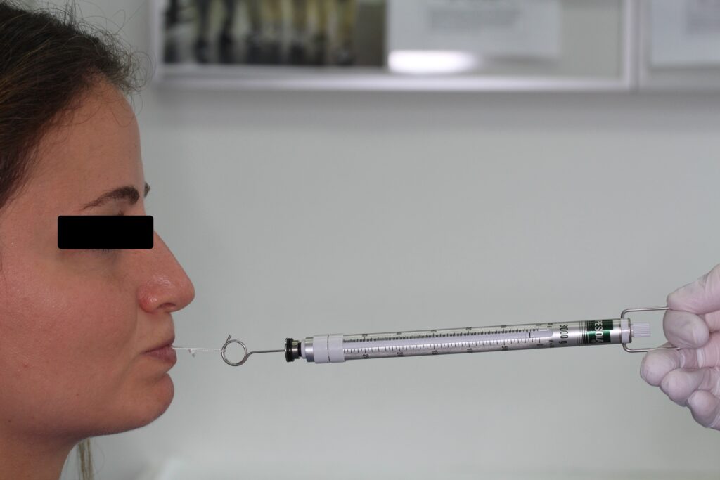
Lip Pressure before, during and after Orthodontic Treatment. A Pilot Study
Hanna Gänzer et al.
The purpose of this study is to determine if fixed orthodontic appliances have an influence on musculature, which in turn has an influence on oral posture.
Lip pressure before orthodontic treatment remains the same as during treatment. There is a decrease in lip pressure during treatment. Lip pressure is also considered as a function of age, sex, and dentition.
The study participants come from the patient pool of the University Department of Orthodontics, who are scheduled for treatment with fixed orthodontic appliances.
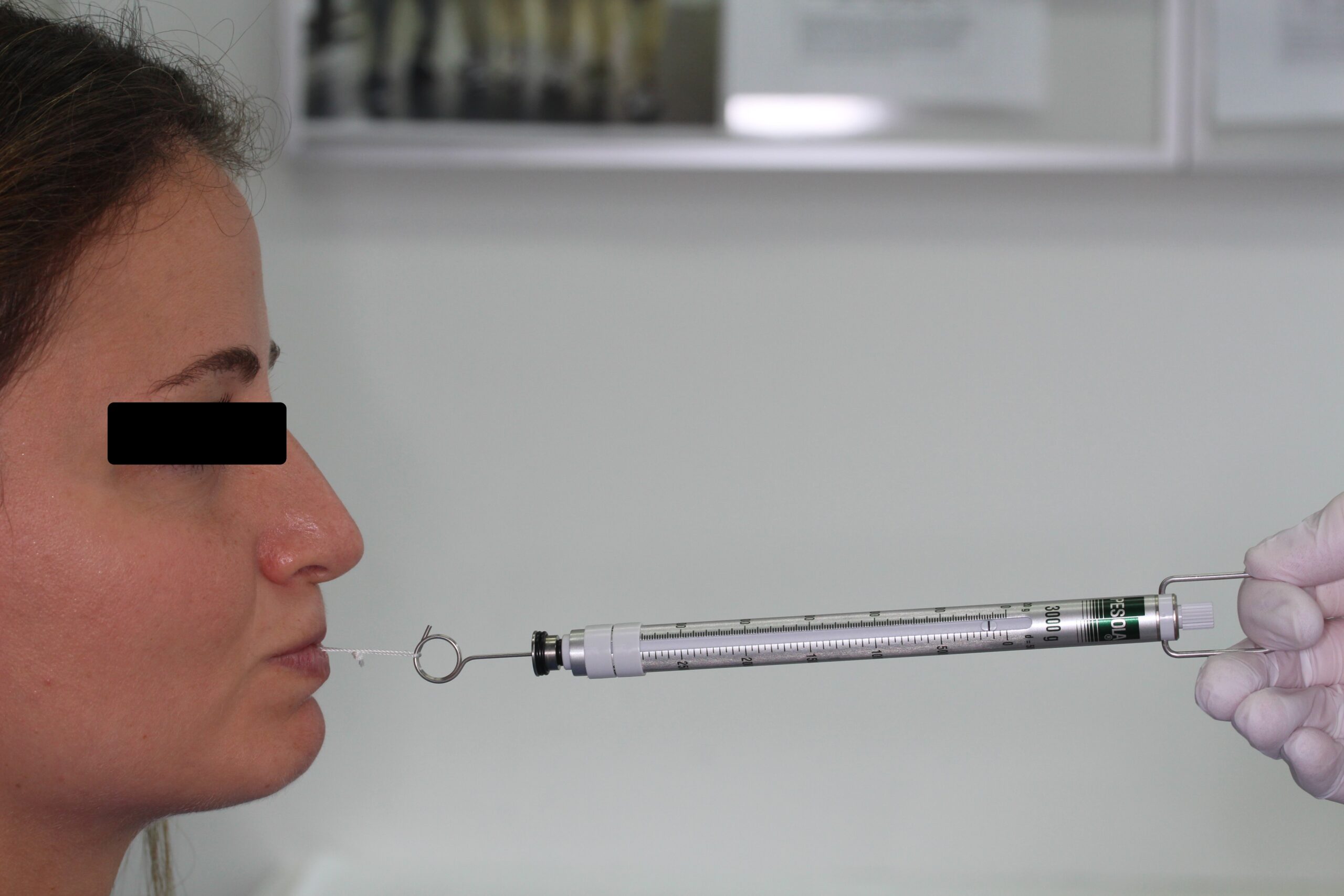
Lip pressure is measured using a myofunctional therapy (MFT) lip scale before treatment begins, on the day braces are placed and after three months (Fig. 7).
A priori, it is expected that there is no significant difference in lip pressure before or during orthodontic treatment.
If, contrary to our expectation, there is a change in lip pressure, it would be interesting to test in further studies whether this change returns to normal after completed treatment and to what extent it affects the position of the teeth. In this case, more frequent prophylaxis could be recommended for patients with fixed orthodontic appliances to counteract the development of gingivitis and periodontitis due to the open mouth posture.
Pictures
Selected Publications
- Riede U, Wai S, Neururer S et al.: Maxillary expansion or contraction and occlusal contact adjustment: effectiveness of current aligner treatment. CLIN ORAL INVEST: 2021; 25: 4671–4679.
- Schieffer L, Manzl C, Schatz C, Haybaeck J, Crismani A: Nrf2 in the Field of Dentistry with Special Attention to NLRP3. ANTIOXIDANTS: 2022; 11: 149.
- Schieffer L, Latzko L, Ulmer H et al.: Comparison between stone and digital cast measurements in mixed dentition. J OROFAC ORTHOP: 2022; 83 (Suppl 1): 75–84.
- Schieffer L, Klawitter T, Ulmer H, Nemec M, Schenz-Spisic N, Crismani A.G: Curve of Spee and Second Mandibular Premolar Agenesis—Present Knowledge and Future Perspectives. APPL SCI: 2022; 12: 11747.
- Artioli I, Crismani A: Das digitale indirekte Kleben von Brackets. INT ORTHOD KIEFEROTHOP: 2022; 54:219-220.
Selection of Funding
- Austrian Society of Orthodontics for supporting the research activities at the University Clinic of Orthodontics
- Tiroler Wissenschaftsförderung (TWF) Dr. Lisa Schieffer MSc. for the project: „ The NLRP3 Inflammasome and Orthodontic Tooth Movement “
Collaborations
- Foss M, Interdisciplinary Nanoscience Center (iNANO), Aarhus University, Aarhus, Denmark
- Erbe C, Department of Orthodontics, University of Mainz, Mainz, Germany
- Gruber R, Competence Center Oral Biology, University Clinic of Dentistry, Medical University of Vienna, Vienna, Austria