
Müllerstr. 59
6020 Innsbruck
Fax: +43 9003 73112
Email: marko.konschake@i-med.ac.at
Website: https://www.i-med.ac.at/Anatomie/
Research year
Research Branch (ÖSTAT Classification)
301102, 301103, 301104, 301106, 301107, 301111, 301112, 301113
Keywords
anatomical collection, Clinical-applied anatomy, digitalisation, human development, immunohistochemistry, lymphology, neuromonitoring, peripheral nerves, pre-/postgraduate education, and ultrasound
Research Focus
Our Institute has 2 major research topics: Clinical-applied anatomy and human development
Clinical-Applied Anatomy
Classical anatomical methods, e.g., macroscopic dissection, are used in combination with modern imaging tools (ultrasonography), operative and biomechanical methods for research purposes always focused on a translational approach. To be able to implement new (minimally invasive) operation techniques and imaging on body-donors-to-science in a high-quality and realistic way, special preservations based on alcohol/glycerine are used.
Human Development
A branch of anatomy, human development is an important mainstay for both, descriptive as well as modern clinical and functional anatomy. This is due to the fact that embryology correlates morphological structures with human development, function and possible dysfunctions as compared to well-established animal models. For our research, human adult and human embryonic samples are examined microscopically. Various histological staining, immunohistochemistry, in-situ hybridisation, proximity ligation assay, light fluorescence and electron microscopy are used and evaluated professionally.
General Facts
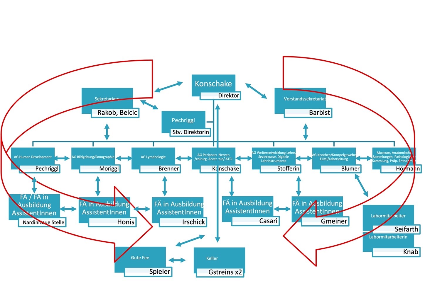
The major tasks of our Institute of Clinical and Functional Anatomy are research and education of human anatomy and embryology. Anatomy aims to understand the normal structures and architecture of the organism and embryology its normal development. Our research and education rely on an extensive body donation program. We offer numerous lectures and pre- and postgraduate courses in anatomy for MD, medical students, PhD students and physicians of different medical disciplines. The main research focus of our 7 research groups always follows a translational, clinically applied approach, with a special emphasis on: Peripheral nerves, human development, imaging, lymphology, skeletal development/ELMI, education and digitalisation and anatomical collection. Therefore, various anatomical techniques are applied in collaborations with national and international universities and university hospitals.
Research
AGs Peripheral Nerves/Surgical Anatomy/ATCI and Imaging/Sonography (Konschake,Moriggl)
All our “Applied Anatomy” studies are carried out with the aim of developing new surgical (minimal-invasive) approaches, improving modern image techniques and new applications for clinical practice. The main applications for clinical and preoperative practice in the field of neuro-sonography are developed on body-donors and volunteers to be able to apply the results to our patients without delay: e.g., in the field of interventional radiology, (plastic) surgery, ENT as well as regional anaesthesia and pain therapy.
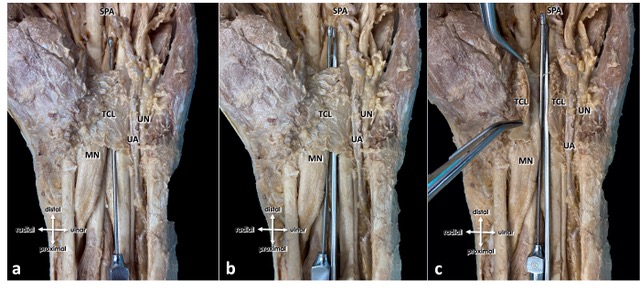
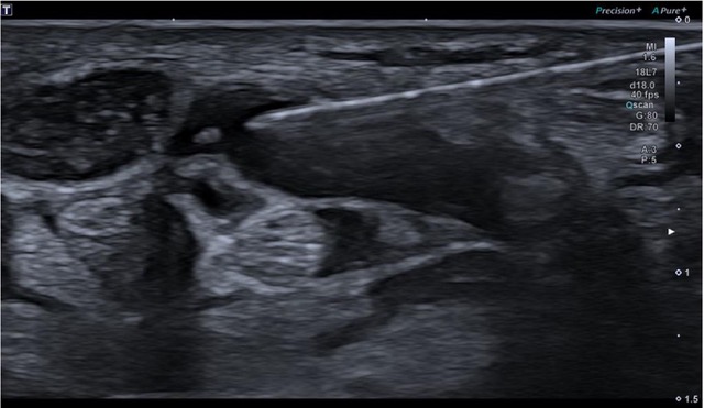
New minimally invasive approaches for various peripheral nerve compression diseases are investigated with the application of ultrasound: e.g., tarsal tunnel syndrome and carpal tunnel syndrome. As a result, patients benefit from a new, minimally invasive surgical method, in which decompression of the peripheral nerves can be achieved via a mere 2mm approach. For example, a new approach of decompression of the median nerve at the carpal tunnel has been developed and evaluated: 104 patients (67 female, 37 male; mean age 60.6 ± 14.3 years, 95% CI 57.9 to 63.4 years) with a clinical and electrophysiological verification of typical carpal tunnel syndrome were referred for a high-resolution ultrasound of the median nerve and were subsequently assigned for an ultrasound-guided CTR. Applying a newly adapted and optimised algorithm was performed using a button tip cannula which has several safety advantages: While serving as a gentle and non-traumatic guiding support for the insertion of the hook-knife, the button tip cannula also doubles as a tool for hydro-inflation. This involves utilizing fluid to progressively expand the operational area throughout the procedure. In all patients, successful release was confirmed by the visualisation of a completely severed transverse carpal ligament during and in the postoperative ultrasound-controls two weeks after intervention. All patients reported a significant reduction of symptoms promptly after this safety-optimised ultrasound-guided minimal invasive CTR. (Fig. 1, Fig. 2)
Another example for our translational approach is the pelvic autonomous plexus. Urinary and faecal incontinence are known complications after low anterior surgical resections (LAR). Preserving autonomous pelvic nerves without any technical assistance is challenging. Therefore, our research investigates the technical feasibility of a new method for pelvic intraoperative neuro-monitoring (pIONM). In twelve female pigs undergoing LAR, the pIONM included direct pelvic nerve stimulation and voltage measurement on the urinary bladder and the rectum for identification of efferent pelvic nerves in the surgical area. Immunohistochemistry of tissue samples obtained after direct nerve stimulation were used for autonomous nerve tissue verification with the aid of S100, VIP, DBH and TH antibodies. In two human body-donors the autonomous pelvic plexus was dissected and the position of the electrode could be displayed. The smooth muscle contraction of the urinary bladder and/or the rectum due to direct stimulation of the innervating nerves is detectable with voltage measurement. The macroscopic contraction of both the urinary bladder and the rectum correlated with a change in voltage drop compared to the status prior to the contraction. Thus, it was possible to identify pelvic nerves in the surgical area.
AG Human Development (Pechriggl)
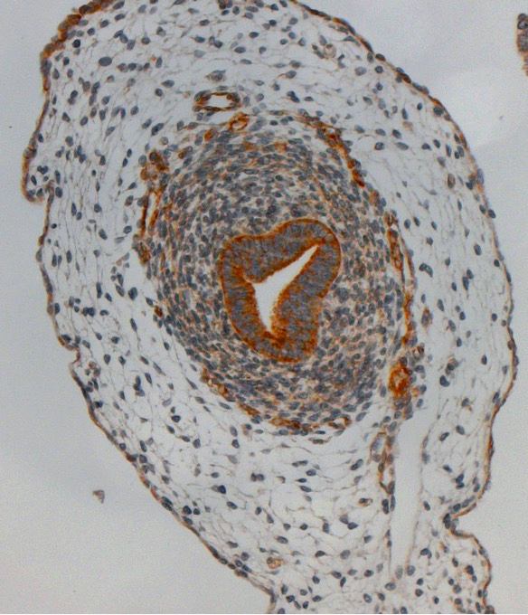
The Human Development group focuses on organogenesis in the late embryonic and early foetal period respectively and its correlation with clinical and functional applications. (Fig.3)
Epithelial ovarian carcinomas are the most dominant and most lethal type with 5 major histological subtypes, which differ in their molecular profile, pathogenesis and prognosis. In the ongoing controversy about the origin of epithelial ovarian carcinomas and especially high-grade serous carcinomas, the organogenesis and differentiation of the various entities is a key component in understanding cancerous dedifferentiation. In humans, despite many animal and cell models, the organogenesis of the human ovary and its organ-specific, sex hormone-independent and dependent developmental programs are still not fully understood. Our aim is to investigate the different stages of early sex development and an assignment of the different epithelial to their embryological origin in human embryonic/foetal preparatons in cooperation with the University Hospital for Gynaecology and Obstetrics Innsbruck.
Therefore, we use histomorphological methods such as immunohistochemistry and in situ hybridisation to demonstrate their temporal-spatial relationships and to make an appropriate assignment to the lateral plate mesoderm and coelomic epithelium of the OSE for patterning and differentiation respectively along the different Mullerian duct derivates.
This may well help toward a better understanding of carcinogenesis will be relevant for the establishment of effective screening tools and novel therapeutic approaches.
Based on our previous research on inner ear development in the early foetal period in cooperation with ENT of MUI, we have been able to expand our field of research in a translational manner and conduct research on age-related hearing loss. For this purpose, we relate senescent changes in human cochlear epithelia and vessels to foetal findings to evaluate the effect of aging and possible influential factors. To evaluate a possible point of application for electrical stimulation, MedEl (MED-EL Elektro-Medizinische Geräte GmbH) could be attained as a cooperation partner.
The AG Human Development cooperates with several partners, both internally as well as externally: organogenesis of adipose tissue (a.o. Prof. Brenner, Clinical and Functional Anatomy MUI), “Dopaminergic neurons in the embryonic brain” (a.o Prof. Wenning, University Hospital for Neurology) and “Fertility Preservation: understanding the effects of low dose ovarian irradiation in a mouse model” (Dr. Elisabeth Reiser, University Hospital for Gynaecology and Obstetrics Innsbruck). Furthermore, our collaboration with Prof. Dr. Beate Brand-Saberi (Institute of Molecular Anatomy and Embryology, Bochum) deals with the genetic investigation of human CNS.
A new elective with regard to the education of natural sciences and medicine students : Stem Cells: Modelling of Development and Disease: Theoretical and Practical Basics in collaboration with Priv. Doz. Irina-Roxana Deleanu (Institute of Neuroanatomy, Innsbruck)
AG Lymphology – Phlebology (Brenner)
The working group deals with those vessels that are directed centrally from the periphery. Lymphatic vessels transport fluid from the extracellular matrix centripetally into the two venous angles; veins transport blood from the capillary bed directly to the heart.
In recent years, a special focus has been placed on book contributions in the field of lymphology and the co-editing of a comprehensive standard work on the subject, which is to be published in 2023.
A disease that is – also – assigned to lymphology is lipohyperplasia dolorosa (LiDo), vulgo lipedema. LiDo (ICD-11: EF02.2; ICD-10, German Modification Version 2022: E88.2-) is a painful disproportionate accumulation of subcutaneous adipose tissue (SAT) in the lower and – less frequently – in the upper extremity of almost exclusive women. The trunk and head are not affected by the disease, nor are feet or hands. Two studies are currently underway in this area of research: (1) the analysis of LiDo patients regarding their Body Mass Index (BMI) and Waist-to-Height Ratio (WHtR), and (2) the analysis of the size of the adipocytes in the affected extremities. In case of the first topic, the monocentric pilot study has already been published (Brenner and Cornely, 2022), and the publication for the multi-centre study is in preparation. Compared to the WHtR, BMI exhibits a general overestimation , which is to be expected. The WHtR does not show a correlation with age, but rather with the morphological stage of the disease. BMI seems to systematically overestimate the cardiovascular and metabolic risk in patients with LiDo. Therefore, in our view, BMI should be replaced by WHtR in diagnosis and follow-up as well as in the planning of comparative studies. The data collection for the topic of adipocyte size has already been completed; evaluation and manuscript preparation are underway. Preliminary analyses suggest that LiDo increases the number of fat cells, but not their size, unlike obesity.
The publications in the field of phlebology concern a broader field. We have mainly dealt with the great saphenous vein, the internal iliac vein as well as the so-called Giacomini vein, and a book contribution concludes this subject area. For the great saphenous vein, we could show that its function can be restored by external valvuloplasty. Furthermore, cellular senescence may have an impact on the development or recurrence of varicose veins. Central venous spurs in the left internal iliac vein in May-Thurner-Syndrome occur solely at the venous confluence of the internal iliac veins (Urbas and Brenner, 2021). They could be remnants of ostial valves, while other types may have other causes.
AG Skeletal Development/ELMI (Blumer)
Skeletal development and colour formation in vertebrates. In vertebrates, most of the skeleton develops from a cartilaginous scaffold that arises from the mesenchyme. The prerequisite for normal bone development is the ingrowth of blood vessels, coupled with the breakdown of cartilage matrix and the formation of bone tissue. These processes cease as soon as the original cartilage is completely replaced by bone. The growth of the bones is thus completed.
Cartilaginous fish (sharks and rays) have a cartilaginous skeleton, which – in contrast to vertebrates – remains cartilaginous throughout life. The cartilaginous core of the skeleton is reinforced by a layer of mineralised tiles, which is enveloped by dense connective tissue (perichondrium). This architecture gives the skeleton both rigidity and elasticity and appears to be the basis for the cartilaginous fishes being an evolutionarily highly successful group.
Cartilaginous fish are usually inconspicuously coloured, but some skate species are distinguished by blue spots on their backs. This colour is not based on pigments, but arises from cellular structures that scatter light and thus produce the blue hue. Therefore, this colour is referred to as a structural colour. The nanostructures responsible for colour production are currently investigated.
Normothermic Liver Preservation and Conditioning. The ultrastructure of the mitochondria is examined in this project.
Pictures
Selected Publications
Loizides A, Honold S, Skalla-Oberherber E, Gruber L, Löscher W, Moriggl B, Konschake M, Gruber H. Ultrasound-Guided Minimal Invasive Carpal Tunnel Release: An Optimized Algorithm. Cardiovasc Intervent Radiol. 2021 Jun;44(6):976-981. doi: 10.1007/s00270-021-02789-2. Epub 2021 Feb 24. PMID: 33629135; PMCID: PMC8172390.
Schuler R, Goos M, Langer A, Meisinger M, Marquardt C, Fritsch H, Konschake M. A new method of intraoperative pelvic neuromonitoring: a preclinical feasibility study in a porcine model. Sci Rep. 2022 Mar 7;12(1):3696. doi: 10.1038/s41598-022-07576-8. PMID: 35256643; PMCID: PMC8901737.
Pechriggl E, Blumer M, Tubbs RS, Olewnik Ł, Konschake M, Fortélny R, Stofferin H, Honis HR, Quinones S, Maranillo E, Sanudo J. Embryology of the Abdominal Wall and Associated Malformations-A Review. Front Surg. 2022 Jul 7;9:891896. doi: 10.3389/fsurg.2022.891896. PMID: 35874129; PMCID: PMC9300894.
Urbas, D., Brenner, E., 2021. Das May-Thurner-Syndrom – der Beckenvenensporn – in heutiger Zeit. Phlebologie 50:184-195. http://dx.doi.org/10.1055/a-1394-3111.
Blumer MJ (2021) Bone tissue and histological and molecular events during development of the longbones. Ann Anat 235:151704
Selection of Funding
- F. 47167/9-2023
- BMBF 13GW0192
- TWF D-155210-015-011
Collaborations
Prof.Dr. José Sanudo, MD, Prof.in Dr.in Teresa Vazquez, MD, Complutense University of Madrid, Center of Body Donation, Madrid, Spain
Prof.Dr. Jeffrey Janis, MD, FACS, Department of Plastic Surgery, Ohio State University Medical Center in Columbus, Ohio, USA
Univ.Prof.Dr. Fabrice Duparc, Université de Rouen, Rouen, France
Prof.Dr. David Chen, MD, FACS, Lichtenstein Amid Hernia Clinic at UCLA Section of Minimally Invasive Surgery, UCLA Division of General Surgery, Los Angeles, USA
Univ.-Prof. Thomas Fichtner-Bendtsen, MD, Ass.-Prof. Jens Børglum Neimann, MD, Department of Clinical Medicine Anesthesiology, Copenhagen, Denmark