
Müllerstraße 44
6020 Innsbruck
Fax: +43 512 900373873
Email: Monika.Ritsch-Marte@i-med.ac.at
Website: https://www.i-med.ac.at/dpmp/bmp/
Research year
Research Branch (ÖSTAT Classification)
103002, 103021, 103037, 103040, 103045, 302044
Keywords
acousto-fluidics, aerosols., biophotonics, computational imaging, diffractive optics, digital holographic microscopy, linear and nonlinear Raman microscopy, optical coherence tomography (OCT), optical tweezers, spatial light modulators, super-resolution microscopy, and UV radiation
Research Focus
The Institute of Biomedical Physics conducts application-oriented basic research projects focused on
developing novel methods and technologies for biomedical research. Currently, there are two distinct
research groups: the Biomedical Optics group, which focuses on advancing optical imaging and
micromanipulation; and the Solar UV Radiation group, which provides data on solar radiation and air
quality.
General Facts
A major research effort of the Biomedical Optics research group is directed towards advancing optical
imaging. Recently, the emphasis has been on adaptive optics in multi-photon fluorescence microscopy
to enable deeper penetration of biological tissue. A second research theme focuses on acoustic and
optical micromanipulation for contact-free handling of microscopic particles.
The Solar UV Radiation Research Group is devoted to optimising spectroradiometric instruments and
developing analysis techniques for solar radiation spectra and aerosol optical depth. The group is
involved in various EU projects concerning ground-based remote sensing and maintains several
instruments for measuring solar irradiance spectroscopically. The group also develops retrieval
techniques for atmospheric air quality parameters. A particular highlight of the group’s activities is the
development and maintenance of the Austrian UV measurement network (UV index).
The Institute maintains a fruitful collaboration with the other institute in the department — the
Institute of Physiology — which is financed by several joint FWF projects on multi-photon deep brain
imaging (PI: Prof. Alexander Jesacher). Further MUI collaborations are connected to the Image-Guided
Diagnosis and Therapy (IGDT) FWF-doc.funds programme.
In October 2024, ERC grantee Bernhard Baumann joined the Institute as a Professor of Biomedical
Optics, covering expertise in the field of optical coherence tomography (OCT). The Institute’s research results have gained a high level of international visibility, as reflected
by a number of prestigious awards and nominations. For example, Monika Ritsch-Marte was
awarded the 2024 Emmy Noether Medal , was nominated as one of 30 finalists for a Breakthrough in
the Physical Sciences at the 2024 Falling Walls Conference, and was elected to the Austrian Academy
of Sciences (ÖAW) and the German Academy Leopoldina.
Research
1) Optical and acousto-fluidic micromanipulation
(Monika Ritsch-Marte, Mia Kvåle Løvmo, Simon Moser, Franziska Strasser, Gregor Thalhammer-
Thurner)
Microscopic particles can be held and moved in a contact-free manner using optical and acoustic force
fields. Our groundbreaking holographic optical force and torque measurement technique offers
unparalleled detail, providing comprehensive information about the forces and torques acting on
particles of any shape, even those with unknown optical properties. This opens up many possibilities
for studying biological specimens, for example investigating cell mechanics. To extend the range of
particle manipulation to larger samples of up to millimetre size, we have also created novel acousto-
fluidic tools in recent years. These tools allow cell clusters, cancer spheroids or organoids to be
reoriented or continuously rotated in a controlled way using actuated MHz ultrasound patterns in a
microfluidic chip. For example, this enabled the first tomographic reconstruction of a five-day-old
zebrafish larva without attenuation artefacts in an FWF-SFB collaboration with Prof. Wolfgang Drexler
at the Medical University of Vienna.

reconstruction of large, optically thick samples such as a zebrafish embryo (published in Nature
Commun. 2024). a) overview of ultrasound-induced reorientation for multi-angle optical coherence
tomography (ULTIMA-OCT), b) artefact-free reconstruction of the sample (comparison with
conventional OCT).
(adapted from publication in Nature Communications (2024))
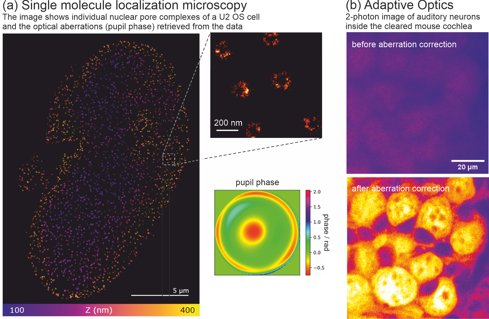
2) Adaptive multi-photon imaging
(Alexander Jesacher, Simon Moser, Juan Muñoz Bolaños, Monika Ritsch-Marte, Maximilian Sohmen, Maria Borozdova, Kibum Nam)
Multiphoton excitation scanning microscopy is a powerful imaging tool that can penetrate deep into
biological tissues. Using infrared excitation wavelengths, which are less susceptible to scattering than
visible light, it can penetrate much further through seemingly opaque tissues, such as the brain and
skin. Our research focuses on significantly increasing imaging depth using adaptive optics (AO)
methods. AO combines wavefront measurements and dynamic spatial beam shaping to correct for
optical aberrations and scattering effects introduced by tissue. Our recent research in this project has
developed better algorithms that can measure aberrations more quickly and reliably. We have also
expanded our set of imaging modalities to include three-photon stimulated techniques, which enable
imaging at depths of up to a millimetre, even in optically dense tissues such as mouse brain.
3) Single molecule localisation microscopy
(Alexander Jesacher, Simon Moser, Julian Maloberti)
Single Molecule Localisation Microscopy (SMLM) is a “nanoscopy” method that enables the
achievement of the highest possible spatial resolution. In collaboration with Prof. Gerhard Schütz’s
group at TU Wien, we are investigating methods to further improve axial optical resolution, in an
attempt to achieve a sub-10 nm accuracy in the z-axis .
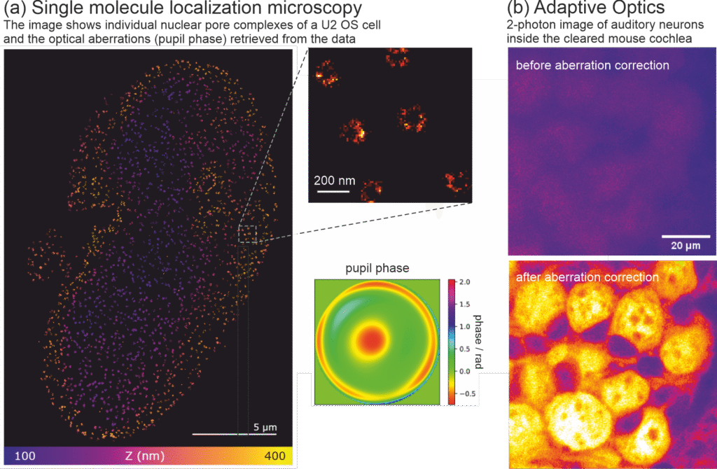
complexes, located at the lower nuclear membrane of a U2 OS cell. The image in
the bottom right shows optical aberrations in the form of a pupil phase image,
retrieved directly from the data using our new algorithm. (b) Two-photon excited
fluorescent images of auditory neurons, located deep inside an optically cleared
mouse cochlea. The images show the cell population before (top) and after
(bottom) correcting for optical aberrations. (collaboration with Prof. Christian Vogl,
Dept. of Physiology, MUI) (Alexander Jesacher, Inst. of Biomedical Physics, MUI; part (a) of the image will be
published soon in Biomedical Optics Express.)
This would enable us to measure the
conformational state of CD3-Zeta, a membrane-bound protein found in T cells, and thus elucidate the
signalling pathways that ultimately lead to the immune response. Recently, we have developed an
approach that allows us to measure optical aberrations directly from molecular signals, thereby
avoiding the systematic errors that often plague SMLM.
4) Tunable lenses and Fourier Optics
(Stefan Bernet, Alexander Jesacher, Monika Ritsch-Marte)
Significant improvements have been made to our patented tunable diffractive lens
technology (also known as Moiré lenses), allowing the focal length to be changed over a wide range at
galvo speed. This is faster than most other existing focal scanning methods. The device has already
been fabricated as part of a Master’s thesis by Matthias Dallio and supervised by Alexander Jesacher.
Additionally, a cost-effective method for high-resolution, high-contrast quantitative phase imaging in
microscopy has been developed by simply integrating a commercially available variable neutral density
filter into the beam path.
5) Solar Radiation and Air Quality
(Axel Kreuter, Regine Gradl, Barbara Klotz, Hanna Rohringer, Michael Schwarzmann, Marie
Stöckhardt, Alma Ubele, Jochen Wagner)
The Solar Radiation and Air Quality research group investigates the implication of solar radiation for
air quality, human health and potential biomedical applications. Within the Austrian UV network, a
successful intercomparison campaign has been performed with the European UV standard QASUME.
The collaboration with the German UV network has intensified through a project involving the real-
time publication of UV index maps of Europe based on satellite data processing.
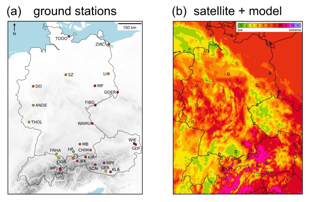
derived UV Index map (b) (published in Klotz et al., Remote Sensing (2025)) (Barbara Klotz, Regine Gradl)
The group continues to operate various solar measuring instruments and sun photometers for aerosol
remote sensing research as part of the Aerosol Robotic Network (AERONET). In 2024, two new spectral
instruments (BTS-UV and BTS-IR) were installed as prototypes for new retrieval tests, particularly for
greenhouse gases in the short-wave infrared spectrum. As part of an ongoing FWF project, a
measurement campaign was performed in Innsbruck in collaboration with the University of Warsaw.
Finally, the group has initiated and is co-leading the Austrian involvement in the pan-European ACTRIS
(Aerosol, Clouds and Trace Gas Research Infrastructure) activities. Notably, the group’s laboratory now
operates an ACTRIS unit for UV-VIS trace gas remote sensing instruments (CREGARS UVVIS-AT).
Participation in ACTRIS is crucial for future research within the atmospheric science community.
Pictures

Selected Publications
1. Muñoz-Bolaños, J. D., Rajaeipour, P., Kummer, K., Kress, M., Ataman, Ç., Ritsch-Marte, M., &
Jesacher, A. (2024). Confocal Raman Microscopy with Adaptive Optics. ACS Photonics, 12(1), 176-
184. DOI: 10.1021/acsphotonics.4c01432
2. Sohmen, M., Borozdova, M., Ritsch-Marte, M., & Jesacher, A. (2024). Complex-valued scatter
compensation in nonlinear microscopy. Physical Review Applied, 22(4), 044036. DOI:
10.1103/PhysRevApplied.22.044036
3. Rothe, S., Wisal, K., Chen, C. W., Ercan, M., Jesacher, A., Stone, A. D., & Cao, H. (2025). Output
beam shaping of a multimode fiber amplifier. Optics Communications, 577, 131405. DOI:
10.1016/j.optcom.2024.131405
4. Moser, S., Jesacher, A. & Ritsch-Marte, M. (2023). Efficient and accurate intensity diffraction
tomography of multiple-scattering samples. Optics Express, 31 (11), 18274-18289. DOI:
10.1364/OE.486296
5. Sohmen, M., Muñoz-Bolaños, J. D., Rajaeipour, P., Ritsch-Marte, M., Ataman, Ç., & Jesacher, A.
(2023). Optofluidic adaptive optics in multi-photon microscopy. Biomedical Optics Express, 14 (4),
1562-1578. DOI: 10.1364/BOE.4814536.
6. Wagner, J., Ubele, A. A., Schenzinger, V., & Kreuter, A. (2024). Extended aerosol optical depth
(AOD) time series analysis in an Alpine valley: a comparative study from 2007 to 2023. Aerosol
Research, 2(1), 153-159. DOI: 10.5194/ar-2-153-2024
7. Klotz, B., Gradl, R., Schenzinger, V., Schwarzmann, M., Schreder, J., Lorenz, S., Gröbner,
J., Hülsen, G., & Kreuter, A. (2025). UV Map Nowcasting and Comparison with Ground-
Based UV Measurements for the DACH Region. Remote Sensing, 17(4), 629. DOI:
10.3390/rs17040629
8. Masoom, A., Kazadzis, S., Valeri, M., Raptis, I. P., Brizzi, G., Papachristopoulou, K., Barnaba, F.,
Casadio, S., Kreuter, A., & Niro, F. (2024). Assessment of the impact of NO2 contribution on aerosol-
optical-depth measurements at several sites worldwide. Atmospheric Measurement Techniques,
17(18), 5525-5549. DOI: 10.5194/amt-17-5525-2024
9. Strasser, F., Ritsch-Marte, M., & Bernet, S. (2025). Quantitative phase imaging with optical
differentiation by spatially variable amplitude filters. Optics Letters, 50(2), 353-356. DOI:
10.1364/OL.540669
10. Kvåle Løvmo, M., Deng, S., Moser, S., Leitgeb, R., Drexler, W., & Ritsch-Marte, M. (2024).
Ultrasound-induced reorientation for multi-angle optical coherence tomography. Nature
Communications, 15(1), 2391. DOI: 0.1038/s41467-024-46506-2
Selection of Funding
1. FWF: SFB F68 “Tomography Across The Scales” (DOI 10.55776/F68), Teilprojekt “Inverse Problems
in Imaging of Trapped Particles” (F6806), Ritsch-Marte Monika, € 540k, 2018-2022 and prolonged
2nd period € 520k, 2022-2026
2. FWF doc.funds: “Image-guided Diagnosis and Therapy” (10.55776/DOC110) Teilprojekt “Infrared
Imaging through tissue by wavefront shaping” (SUB05) Ritsch-Marte Monika, € 190k, 2021-2026
3. FWF: „Deep 3-photon brain imaging with wide scatter compensation“ (DOI 10.55776/P36687)
4. Alexander Jesacher, € 609k, 2023-2027
5. FWF: “Live Cell Superresolution Imaging of Protein Conformation” (DOI 10.55776/P36022)
6. Gerhard Schütz (Co-PI Alexander Jesacher), € 396k, 2022-2026
7. FWF: „Deep brain vision: 3D adaptive 2-photon microscopy“ (DOI 10.55776/P32146)
8. Alexander Jesacher, € 489k, 2019-2024
9. FWF: “Synergetische Aerosol Fernerkundung mit Pandora in ACTRIS“ (DOI 10.55776/I6016)
Kreuter Axel, € 334k, 2022-2026
10. Bundesministerium f. Nachhaltigkeit und Tourismus: „Messung und Analyse der solaren UV-
Strahlung in Österreich“ (D-150200-017-012) Kreuter Axel, € 1.300k, 2019-2029
11. EU: „ACTRIS ERIC“ (D-150200-017-015) Kreuter Axel, € 172k, 2023-2028
12. EU: „Grasp Synergy“ (D-150200-017-016) Kreuter Axel, € 73k, 2023-2027
13. EU: “ATMO ACCESS” (D-150200-012-014) Wagner Jochen, € 75k, 2021-2025 (prolonged)
14. EU: “ACTRIS IMP” (D-150200-012-012) Wagner Jochen, € 66k, 2020-2023
Collaborations
Martin Booth and Patrick Salter, Univ. of Oxford, Oxford, UK
Hui Cao, Yale Univ., New Haven, USA
Schmidt, Michael, Friedrich-Alexander University of Erlangen-Nürnberg, Erlangen, Germany
Wolfgang Drexler, Medical University of Vienna, Vienna, Austria
Schütz, Gerhard, Technical University of Vienna, Vienna, Austria
Çaglar Ataman, Univ. of Freiburg, Freiburg, Germany
Alexander Cede, LuftBlick OG, Innsbruck, Austria
Sebastian Lorenz, Bundesamt f. Strahlenschutz, Neuherberg, Germany
Bruce Drinkwater, Univ. of Bristol, Bristol, UK
Halina Rubinsztein-Dunlop, U. of Queensland, Brisbane, Australia